


This talk was first given at the American Osteopathic Association of Orthopedics (AOAO) this month at their annual spring meeting. It discusses risk factors for persistent pain after hip arthroscopy and a decision tree for management.
Transcript
4,555 words · 23 min
Transcript
4,555 words · 23 min
Good afternoon everyone. I wanna thank you for joining me and allowing me to speak a little bit about some advancements in Hip arthroscopy and in particular, what are we doing differently with our decision tree for when patients come in with a failed Hip arthroscopy or persistent Pain.
I have no relevant disclosures for this talk and we're gonna talk about a couple of key points here. The first being, where are we in 2021 with Hip arthroscopy?
Identifying who's an at-risk surgeon for potential complications or failures. We also identify the appropriate patients to operate on, and more importantly, who's at risk of failing in that patient population. And then finally, we'll, we'll summarize with the decision tree on how we're moving forward with failures at this point.
So Hip arthroscopy is growing. It's up over 600% in the last 10 years. Uh, I had the opportunity to lecture, uh, at many fellows courses, and I can tell you from just 10 years ago when I was a fellow, uh, to just this current year, the number of fellows being exposed to Hip Arthritis.
Earthroscopy and planning to do it in their practice, uh, is exponential. And, and why is this? Well, number one, sports fellowships are now incorporating this, and Hip arthroscopy is considered, uh, not just a niche anymore, it's actually considered something that should be part of sports fellowships. And also, there's Hip preservation fellowships that are out there. There's 5 dedicated ones, uh, that are known right now, and many others that are unaccredited. And in the industry setting, we know Hip arthroscopy is one of the fastest growing markets that there is.
So, what do we know? Well, there's dedicated advancements, uh, that's been made over the last 20 years since his Hip arthroscopy was first described in 1999. And really, we found that there's instruments now that are designed specifically for Hip arthroscopy that are longer and, and tapered for the uh femoral head. We know that anchors have been reconfigured to allow for shorter anchors as well as not less configurations that allow for us to better repairs.
There's Hip arthroscopy tables, uh, there are some that are traditional with posts, some that are postless, some that are supine, some that are lateral, so there's a, a variety for all of our surgeons, and the number of teaching courses in particular, whether it's one day courses, weekend courses, or even many fellowships, has certainly risen over the last several years, both in industry as well as to our societies.
But the question still remains with all of this, are we training physicians accurately to be proficient in Hip preservation?
So who's an at-risk surgeon? Well, if you go through the literature, there's many of us that are. There was an ACGME study that was performed in 2015, 2016, looking at the residents that were graduating from orthopedic residencies in the US. They found that over a five-year period of residency, the average orthopedic resident was graduating with about 8 to 18 Hip exposures for arthroscopy, with the median being 5 Hip arthroscopies over the course of a full 5-year training program.
And, and part of this is we know there's only 5 dedicated Hip preservation fellowships out there, and if we weren't producing a Hip arthroscopists that were in the community, many of these residents weren't seeing this, but maybe that tide is gonna change.
What we do know is this, Ben Dome and his colleagues looked at what it takes to become proficient with Hip arthroscopy, and the numbers are a bit surprising. So to access the Hip joint, just to get into the case and get started, it takes almost 20 cases to become proficient.
To perform a labor repair accurately and safely, it takes about 34 cases.
To do a bony resection, acetabuioplasty, or femoral osteoplasty, 54 cases. To perform a labral reconstruction, most commonly seen with revision Hip arthroscopies, about 100 cases. And to perform a capsular closure at the end of a routine Hip arthroscopy or application for someone with instability, a little over 50 cases.
So when you look at this a little further, they actually found that when you look at Hip preservation fellowships, they recommend 128 cases be performed to become proficient coming out of a one-year fellowship. And part of this is that Hip preservation also entails not just arthroscopy like there is in sports fellowships, but also open cases as well. And in sports fellowships, they found that 67 cases was required to become proficient coming out of fellowship.
They also said that about 56% of the surgeons surveyed for this study, they felt that you should not change gears, or you should not become a Hip arthroscopist midway through your career. Meaning if you haven't been doing this coming out of fellowship, the odds of you being able to pick this up midway through your career and be proficient at it, were pretty low in their opinion, and perhaps it's something you shouldn't adopt.
So this study by Lee and Ku also looked at complications and surgical time proficiency. And what they found was, after about 30 cases, things began to level off and plateau, regarding the number of complications you would see, as well as the surgical time for each case. So think about that, 30 cases it would take, and if you're not doing a lot of Hip arthroscopy, that's something that can actually lead to quite a long time, maybe several years before you hit that 30 case margin.
So why is this? Why is this different than Knee arthroscopy and Shoulder arthroscopy? Well, it's technically demanding, and the first purpose of that is we're using a seventy-degree arthroscope.
Next thing to consider is the hips is a deep joint. It's not superficial like a Knee or a Shoulder. Even in a Shoulder which is muscular, it's a relatively small space to get into, whereas the Hip is a much deeper uh place to be.
And there's multiple bits of Pathology that can be uh sought after. Some of these are labral tears, some of them are bony impingement known as FAI, loose bodies, micro instability. This can be due to capsule laxity or ligamentum tears. So again, depending on what you're going after as a surgeon for this Pathology dictates how much proficiency you need, going back to the earlier Ben Do article.
So we also know the Hip arthroscopy has failures, and, and why is this? Well, some of it's just inappropriate management of the Pathology, and this could be due to a lack of knowledge, understanding what's normal versus abnormal, what's at risk for failure. Some of it can be due to a lack of inappropriate imaging. Revision cases, in my opinion, should undergo a CAT scan to look for bony deficiencies and bony abnormalities.
Some of it's just a lack of technical skill. It's very difficult to use a 70 degree arthroscope, let alone one in a deep space like the Hip joint.
And really the thing that's most striking is, you know, inadequate addressment of all pathologies. So if you're not comfortable doing a bony resection on the acetabulum, or if you're uncomfortable doing a large cam resection on the femoral neck, now this is a problem, this can lead to failures down the road. But ultimately, the number one failure with any Surgery is Patient Selection.
So FAI, we know that FAI is certainly the the picture of Pathology seen above here, and this could be in one of any number of shapes and sizes. Picture A shows a typical Hip where there's no anatomic variants. Picture B demonstrates a femoral cam lesion where you get this bit of a humpback appearance on the femoral head and neck, which will allow for crushing of the uh the labrum as the Hip flexes up into the acetabulum. Picture C demonstrates a pincer lesion or an overhang of the acetabulum.
Again, this could be a bit of a bird's claw that comes down and it leads to maceration of the labrum with repetitive flexion. Then picture D, which is the most common, is a mixed Pathology of both an acid tab overhang, as well as a cam lesion.
So going to the basics, imaging is the most important, and you can't get away from getting good radiographs to start off. So in my office and in many offices, AP pelvis and a dedicated Hip view are important, and you want to make sure for your dedicated Hip view, you're getting a 20 degree internal rotation on the pelvis, because this will help give you your best crossover sign evaluation for the pincer lesions.
For the lateral X-ray, I prefer a 45 degree done view to help bring this femoral cam lesion into view, to allow for measurement of the alpha angle as seen in this picture below.
And then advanced imaging should be used judiciously. CAT scans are reserved in my hands for disc revision cases. MRI, arthrogram, and direct MRI and indirect MRI are also used at time of uh decision for looking for a Labral Tear.
When you start looking at your plane radiographs, so, again, you can get the most bang for your buck just off of simple measurements with appropriate radiographs. For a cam lesion, an alpha angle is going to be measured at 50 to 60 degrees. It's important to know how to measure this alpha angle on your own. The picture on the left depicts a line dropped down the center of the uh the femoral neck into the center of the femoral head, and then there's a transsectional line coming out laterally here, just at the superior margin of that femoral cam lesion.
Again, over 50 to 60 degrees is going to be indicative of a femoral cam lesion. For a pincer lesion, you're gonna see a lateral center edge angle of greater than 40 degrees, and for a mixed component, you'll see a variety of these as well.
So what sort of Approach do we take to patients that failed Hip arthroscopy for labral tears? Well, Shaneno and his group looked at this and found that failures are multifactorial, and in fact that these are patient related, these are surgeon related, and oftentimes they can be post-op related if there's inappropriate Rehabilitation, or the patient's noncompliant.
So the number one thing that they found in their study was simply we have residual bony deformity, meaning we had inadequate resection of our femoral cam or a pincer lesion, and this led to failures and persistent Pain.
The second most common thing they found was capsular deficiency. This could be due to inadequate closure of the capsule, or it could be due to failure of the capsule closure. Remember, if you're gonna do a T capsulotomy for a femoral neck large resection of the cam lesion, you need to close at least a side by side vertical incision for that T, or you're gonna have micro instability that can lead to further complications.
Another study that was done by Chris Larson and his group looked at this, and again, they found the most common reason for Revision Hip Arthroscopy was uh remaining bone or incomplete resection and correction of the bony abnormalities on the femur and the the acetabulum. In their study looking at 50 patients, they found 90% of the patients, or 45 of the 50, had residual bone that was malcorrected and required further surgical intervention.
So why is this? Is it that we're positioning the patients inappropriately, being careless, not getting true positioning during surgical intervention?
Is it that patients are just failing to be recognized for their deformity by the surgeon? Or is it a comfort level thing where the surgeon is just not comfortable resecting that bone off the the acetabulum or the femur?
Well, who's at risk? Well, we know that, you know, every patient comes into us with an expectation and a goal of being 100%. But the reality of this is we need to weigh our expectations of our patients with what we can comfortably give our patients, taking into all accounts such as their age, their BMI, the amount of the tear that they have, and the amount of Arthritis that they have. Expectation equals outcome. And the literature suggests inferior results with people that have pre-existing Osteoarthritis, workman's compensation, and elevated BMI, and smokers.
Some Other residuals include diabetics and non-compliant patients, particularly with postoperative therapy regimens.
So overlapping Pathology could be the most confusing, and it's important not to get tunnel vision just on that MRI that says there's a Labral Tear. We need to remember that patients have Pathology both within the joint, as well as in the peritrochanteric space, as well as the abdomen, and one of the most commonly seen things that uh seen things that we will see with a Labral Tear, particularly in an athlete, is a sports hernia.
Athletic pubalgia cannot be missed, and something that's oftentimes addressed in my operating room and in many operating rooms, in conjunction with the general surgeon at the time of the labor repair Surgery.
And we need to remember that a lot of the Pathology that we see in the Hip can also be attributed to the back. So doing a good back workup, especially if someone's having radicular symptoms, is important because there's nothing worse than doing a great Surgery and having the patient tell us they're still the same symptoms as when they presented.
Recognition of Hip dysplasia is another very, very key important factor that we need to keep in mind. We know that there's been several articles written over the years looking at Hip dysplasia and Hip arthroscopy. At the end of the day, the results are all very poor, and the ultimate outcome is this, if you don't address the malalignment, particularly the fact that there's a small center edge angle of the acetabulum, there's a high probability of a retear, failure, and or acceleration of the Osteoarthritis leading to a rapid turn into a Hip Replacement.
So, this was a study done out of the Iowa Journal of Orthopedics by Barron and Dutchman, and what they found was they looked at about 785 patients uh through a registry, and they looked to see really is the failure rate as bad as we think that it is. And in their study, they broke this down into male and female, age under 40, age over 40. What they found was they had about a 70% failure rate for patients over the age of 40, and they had an overall failure rate of 18% at all comers.
When they broke this down a little further, they found the highest odds ratios for failure were things you would expect. Underlying Osteoarthritis, age less than 40, not that they thought that they were gonna fail, but they had a high risk of Revision surgery. Diabetics and smokers also had a higher rate of failure as well. And interestingly, psychiatric comorbidities. These were also things that were independent, but also true and something to note in your patients when you consider doing Hip arthroscopy.
So the Osteoarthritis is probably one of the biggest things that that puts us on the edge because Osteoarthritis can be subjective, even when using classifications that are standard such as the tones score, and really the gray zones existed between Tonus 2 and Tonus 3, and figuring out the patient is a good candidate or for stretching our indications.
So Larson and Taylor looked at this in, in a two group sort of cohort. They looked at patients at FAI with Osteoarthritis, looking at them with Tonus 2 and Tonus 3 scores, and patients without Osteoarthritis with FAI. What they found was if you had a tonus score grade 2 or more, or less than 2 millimeters of joint space remaining, there was a 52% failure rate in that group, even if you corrected the femoralrostabular impingement. So 1 out of every 2 patients failed in this group.
The recommendation was to avoid Hip arthroscopy in patients with a tonus score of 2 or more, or joint space of less than 2 millimeters. But Tom Bird refuted this a little bit. In his study in 2018, he actually found that tonus scores grade 2 or more, excuse me, grade 2 can still have meaningful positive outcomes with appropriate patient expectation management. They found that age and sex were not positively correlated to this.
So again, there is a gray zone and there's no consensus on this, but again, if you have a patient with a TNA score of grade 2, there has to be a very finite conversation between you and the patient, indicating what you think their outcomes can be, what their expectations should be going forward.
When you look at the best practice for Hip arthroscopy, they surveyed a group of surgeons that were high volume users, and they found some consensus and some not. Only 59% agreed that a post-operative brace should be used. I can tell you my own practice, we don't use the brace for many reasons, but most importantly, most of the insurances are not covering it in my area, so I did not want to have two separate Protocols for my patients.
The group that was surveyed here found that 100% of them were were performing labor repair, and this differs from that earlier study we looked at from the Journal of Iowa Orthopedics with Dutchman, where the most commonly performed labor work was actually labor resection or debridement.
All the surgeons in this study prescribed HO prophylaxis, and 90% of them were performing capsule repair, which is not the standard through the rest of the country. This led to an editorial that was done by Dean Matsuka, looking at should we repair the capsule or not, and, and really there, there is no consensus, but really more a thought process on who should be repaired and who should not be repaired. The repair group should always be considered in a high-level athlete, a dancer, or someone who's hypermobile.
And you want to think twice about repairing the capsule in someone who's arthritic, as this may lead to over tightening of the joint.
So what do we do with a patient that has persistent Pain and failure and presents to your office? Well, first things first is to go back to square one. We need a thorough history. We need to redo our clinical examination, and we need fresh radiographs that you take and make sure are accurate.
I have my go to moves for my physical examination. I, I prefer to do a McCarthy's test, looking for a labral Pathology, and I wanna reproduce the symptoms that the patient is experiencing. Again, impingement, I wanna make sure that I'm not lying to myself. I wanna make sure that I have a good assessment on both hips for where their symptoms are with impingement testing.
It's important to again go back to the location, make sure that they're having Pain in the area they're describing, and also rule out Other areas of Pathology. There's great mimickers, as I mentioned before, with athletic pubalgia, as well as gluteus medius tears, and again, make sure we're not missing any sort of Lumbar radiculopathy.
So this sort of diagram was put together by Ben Do, but it, but it's important, I think really sums up the entire crux of this.
With a patient has failed Hip arthroscopy, you need to have a very concise decision tree. And the way they explained it was this, if they come in with a failed Hip arthroscopy, you need to rule out intra-articular Pathology versus extraarticular Pathology. So they're gonna opt for a diagnostic Injection. The patient responds to the diagnostic Injection positively, we need to go back and assess, all right, is this due to Arthritis, and are we missing this, so we're just covering up the Arthritis with the Injection, or is there actually a Labral Tear going on?
If we determine this is arthritic in nature, we need to have a conversation about non-operative measures such as therapy and and palliative injections as we buy time, or consider Hip Arthroplasty.
If the patient had a negative Injection, we need to look for an extra-articular workup such as gluteus medius tears or Lumbar radiculopathy.
Assuming the patient responds positively to the Injection, you feel as though based on your exam, there's a retear of the labrum, we need to do a very good careful analysis as we move towards Hip preservation revision. We need to understand is there instability of the patient, micro instability due to capsule laxity, or is there again a recurrent Labral Tear or bony uh leftover or bony undercorrection. If there is bony undercorrection, we need to consider doing a revision resection of that bone and or a revision labral repair.
If there's capsule laxity, we need to determine was there a previous repair done by yourself or the uh prior surgeon. We need to be prepared to go and revise the capsule and or augment it with either a graft or xenograft.
Once we make this decision tree possible, we can move forward to the operating room, but rarely should you move forward to the operating room without a clear picture.
I can tell you in my hands, as well as in this Venn diagram that's here, we need to make sure that if we're gonna go back for a labral Revision surgery, the patient's consented for a labral reconstruction because oftentimes the labrum is atrophic when you get into the joint, macerated, or not salvageable. And you need to make sure that you're proficient at doing the labral reconstruction. You don't want to send the patient back for a third Surgery to a different surgeon if you're not comfortable doing this.
So a word of caution on repeating MRIs and MRI arthrograms. You gotta be careful because the MRI's are really, really inconspicuous sometimes in identifying a re-tear. This came out of the Journal of Skeletal Radilogy, and they found that really the best way to identify a labral retear is with arthroscopy, as MRIs can sometimes show post-surgical changes.
This picture here also shows another reason why MRI can be unreliable. While the MRI may show that the Labral Tear is intact, you can see here you've lost your suction seal as this was over tensioned, and the labrum was peeled back, almost like a lip being peeled back by a small child. And again, this loss of suction seal is going to lead to failure down the road or micro instability.
So let's think outside the box for a second. I, I, I love the Needle arthroscopy, and in my hands, if I think someone has the possibility of incompetency of the, uh, the labrum and or the capsule, or a retear, I'm gonna think about doing a Needle arthroscopy for a couple of reasons. One, I can evaluate.
To tear. I can evaluate impingement with flexion and dynamic internal rotation. I can evaluate for loose bodies and capsular deficiency, and I can also deliver a therapeutic Injection at the conclusion of that test. That gives me a lot of information and a lot more real-time information than an MRI. But again, limitations include you can't evaluate the periotrochanteric space, and you can't see all the way into the acetabule.
So I'm gonna leave you with the case that I had of a patient had a previous Surgery performed by a surgeon, not me. Uh, I did not get the operative report as it was done out of state, but they had persistent Pain with symptoms and the MRI was indeterminate, so I opted to do a diagnostic Needle arthroscopy. Again, here, I marked out my center of my joint, uh, equal with the top of the trochanter, and I marked out my traditional mid anterior portal and a lower mid anterior portal.
What we're able to do here was we were able to identify the patient as you access to the mid anterior portal, I'm able to use a little bit of fluid to blow the capsule out of the way. I can see the labrum. You can see there's a red blush on the labrum as well as some adhesions of the capsule to the roof of the acetabulum.
As I begin to dynamically evaluate the Hip, I can see there is some labral insufficiency and some labral tearing, that could be leading to some pinching. And more importantly, as my assistant begins to flex the Hip up, I'm able to see there's some residual cam that's there, that is causing some impingement upon that labrum. This will come into view in a second as we rotate the Hip up and internal, and as we pull back a little bit, you can see there's that cam lesion there with a bare spot on the bone, indicative again of a labral sort of impingement Pathology.
So again, decision making, think about Physical therapy, make sure it's done right. Speak to your therapist to ensure the patient has done an adequate job of therapy. Manage patient expectations. Not everyone can be fixed, and some people are what they are. We need to understand that and explain that. Get a second opinion from a colleague, not some random stranger, but someone that you trust, someone that won't throw you under the bus, and someone that will give you an honest opinion. Consider orthotics if there's any sort of low back Pain or any sort of scoliotic curve the patient may have.
A good Spine evaluation and pelvic floor evaluation and pubic, uh, bone and, uh, sports hernia evaluation is important, and consider injections, both diagnostic and therapeutic, be it steroid or biologic in nature.
Again, be prepared to do things that you may not be comfortable doing, and if you can't do it, get it to the right person. Capsule reconstruction and repair is sometimes paramount and needs to be undertaken. We don't want to leave this patient again without a capsule, and again, the labor reconstruction is something you should be proficient at before going back into the Hip for Revision surgery.
Ultimately, some people are gonna end up with a total Hip. We need to make sure that you're not the cause of it. Be very careful with dysplastic hips. Don't try to fix a Labral Tear in a dysplastic Hip as you're going to accelerate the risk of Arthritis and failure.
Overall, we can prevent many things. We can do a better job of training our younger surgeons coming out of practice. We can all use a very concise decision tree for algorithms. We need to evaluate our patient Risk factors. We need to evaluate our own intrinsic Risk factors, and again, consider reasons for revision only that are correctable.
Under resection of bone on the first time around, a repair of symptomatic labrum that's there, labral incompetency, evaluate previous arthroscopic pictures if you can to see what the labrum looked like at the time of repair. And again, if the capsular repair wasn't done the first time around, consider a repair or a reconstruction.
Thank you so much for your time and attention. I hope this was helpful for you in your practice.
Automatically transcribed and not checked by a person — it may contain errors, particularly in names, drugs and dosages. The recording is the source.
Check yourself
5 questions5 questions on what was actually said in this recording, and we remember how you did. Sign in to take it — an account is free, and nothing else on doc.social asks for one.
AI-generated from this recording’s transcript, not written by the speaker. Tell us if a question misreads what was said.
Related lectures
 Lecture
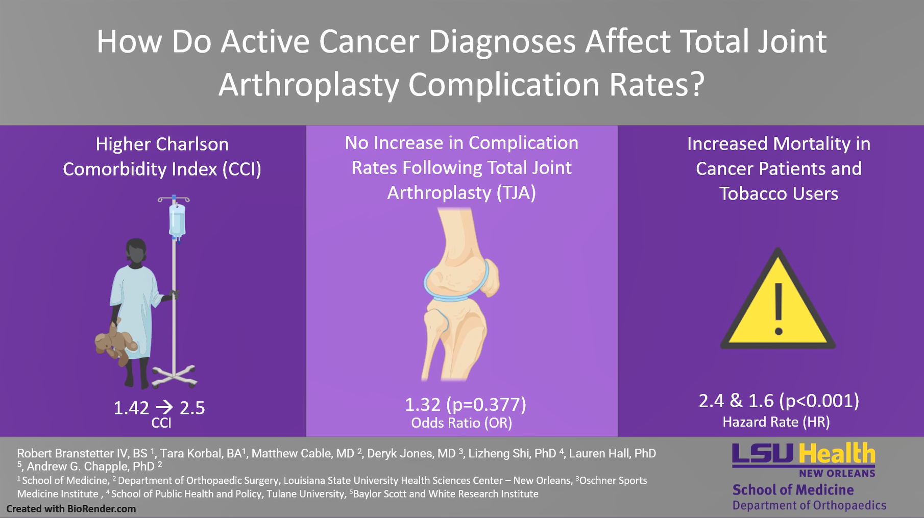
How Do Active Cancer Diagnoses Affect Total Joint Arthroplasty Complication Rates?
There is currently a paucity of data analyzing total joint arthroplasty (TJA) disparities among patients with active cancer diagnoses.
Lecture
Targeted Nutrition in Orthopedic Surgery - A Missed Opportunity to Enhance Outcomes
Surgery triggers a series of inflammatory, immune, and metabolic responses. The stress response leads to an increase in protein breakdown that outpaces protein synthesis. The body's natural store of essential amino acids exit skeletal muscl…
 Lecture
Rotator Cuff Repair with Biobrace augmentation and Bone marrow concentrate
Take a look at how to increase your success rate for healing a torn rotator cuff!